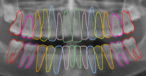
In dentistry, imaging data (X-rays) is the first source of information for an initial assessment of a patient’s dental health and is also the basis for further planning of a patient’s treatment. Both extraoral images, such as a panoramic radiograph or a lateral teleradiograph, and intraoral images, such as bitewing radiographs, are used. Panoramic radiographs are typically the first image since all of the teeth, including roots, are pictured very clearly.
More extensive analyses cost time
First, doctors need to visually analyze the features detectable in the images (cavities, fillings, etc.) and then document them. More extensive analytical methods, such as determining angles and ratios between landmarks in lateral teleradiographs that must be manually identified require significantly even more time and effort.
Automatically extracted information supports medical staff
The Visual Healthcare Technologies Competence Center is working on solutions in this field that make it possible to automatically analyze the imaging data and present the extracted information to doctors. The fundamental first step for this is to automatically detect the contours of each tooth in the images. To accomplish this, the domain-specific knowledge of experts is reproduced by a statistical model that maps both the relative spatial relationships and the variance in shape of the teeth. Using deep learning, this model is initially placed on an image. Then the teeth in the image can be identified and their contours detected.
The final segmentations of the individual teeth can now be used to find individual features of these teeth and output them collectively.
 Fraunhofer Institute for Computer Graphics Research IGD
