 Fraunhofer Institute for Computer Graphics Research IGD
Smart pulmonary ultrasound for COVID-19
POCUS4Covid19 is a new type of software designed to make it easier for doctors to monitor a course of COVID-19 in a patient. To do this, the software automatically analyzes predefined indicators in ultrasound images of the lungs, making an evaluation easier. A special module prevents third parties from using the collected imaging data for purposes other than those for which consent has been given. POCUS4Covid19 is a joint project by the Fraunhofer institutes IGD and IOSB.
(Darmstadt/Karlsruhe) At the start of the coronavirus pandemic, the demand for reliable diagnostic tools in outpatient clinics, ICUs, and other hospital settings swiftly grew. Aside from CT scans and X-rays, thoracic ultrasound proved a suitable instrument for diagnosing viral lung infections induced by SARS-CoV-2. Thoracic ultrasound involves no radiation and can be readily employed at the hospital bed as many times as desired¾and especially makes an interesting option for point-of-care ultrasound (POCUS). The probe is typically the only thing to come in contact with the patient, allowing the equipment to be disinfected easily after use.
Old: visual image analysis
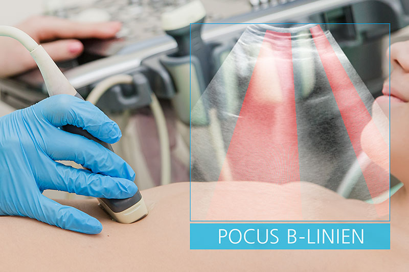
Conventional ultrasound (U/S) is based on the purely visual analysis by a doctor of a currently displayed image. With pulmonary ultrasound, there are clearly identifiable patterns that indicate viral pneumonia. Changes in the imaging data allow the doctor to evaluate the course of the disease. Nevertheless, pulmonary U/S imaging data is not so easy to evaluate and not every examiner is specifically trained in pulmonary ultrasound. However, especially in the current pandemic situation, a quick and clear evaluation of U/S imaging data is vital.
New: POCUS4Covid19 -- software for evaluating COVID-19 pneumonia
This is where POCUS4Covid19 comes in: Using quantitative, computer-assisted image analysis, objective parameters are derived from the U/S imaging data that are made interpretable and useful in evaluating the course of treatment. This software solution provides direct benefits to the acute treatment of patients. The analysis of the U/S imaging data collected over the course of treatment also makes it possible to learn from the initial wave of infections in order to be as prepared as possible for any subsequent waves.
Automatic analysis of characteristic COVID-19 indicators
To this end, the Fraunhofer Institute for Computer Graphics Research IGD continues to develop software that specifies existing scanline algorithms for U/S imaging data, allowing characteristic indicators of COVID-19 to be automatically detected. For example, the software analyzes what are called B-lines, which reflect the interstitial edema found in viral pneumonia. The specific goal is to automatically detect these B-lines in the imaging data and to quantify their value. Other characteristic indicators were established by DEGUM, the German Society for Ultrasound in Medicine, in a COVID-19 protocol that serve as a basis for future updates to the POCUS4Covid19 software.
Secure access to patient data
At the same time, access to the collected clinical U/S data will be made easier and standardized. Typically, imaging data is stored on the ultrasound device itself. Fraunhofer IGD is planning separate software with an interface that allows access to this U/S imaging data via the Medical Data Space (MeDS). The Fraunhofer Institute of Optronics, System Technologies and Image Exploitation IOSB is developing a connector for linking the POCUS software to the MeDS. This interface is designed for two types of U/S imaging data: still images and cine images, or video streams. The MeDS offers a trusted infrastructure for the secure exchange of health data between various parties, such as patients, doctors, hospitals, service providers, and manufacturers. Use of the data can be managed in a user-friendly, transparent, and GDPR-compliant manner. Modern algorithms help with the targeted use of the data.
Unprecedented development
Until now, the computer-assisted analysis of U/S imaging data for COVID-19 diagnosis and treatment has led a shadowy existence. For one, stored U/S imaging data is not as structured or accessible as CT imaging data with its PACS servers. For another, there has yet to be an automated algorithm specifically for lung analysis. Both aspects are being addressed in this project. As part of the “Fraunhofer Solution Days”, a joint talk will provide insight into how the MeDS can be used to securely transfer imaging data in compliance with data protection laws, using a POCUS diagnosis of COVID-19 as an example.
By the image analysis experts
The automatic analysis of U/S images is one of the core competences of Fraunhofer IGD. Since the start of the 2000s, the institute has been developing new applications for ultrasonography, including state-of-the-art methods for automatically processing, analyzing, and integrating medical imaging data. This allows a plethora of essential, clinically relevant information to be extracted and the imaging data to be optimally used along the entire treatment chain.
By the experts for secure data utilization and processing
The “Interactive Analysis and Diagnosis” department develops assistance systems and intelligent, interactive environments. The design method used, Privacy by Design, ensures that data protection laws are seamlessly integrated when developing new technologies.
Further information:
- More about point-of-care ultrasound at Fraunhofer IGD (igd.fraunhofer.de)