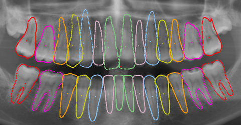
In the dental field, image data (X-ray images) represent the initial source of information for a first assessment of a patient's health status and also serve as the basis for further planning of the treatment process. Both extraoral images, such as the orthopantomogram (OPG) or lateral cephalometric radiographs (LCR), and intraoral images, such as bitewing radiographs, are used. The OPG is the typical initial image because all teeth including roots are clearly visible.
More comprehensive analyses take time
The features recognizable in the images (caries, fillings, etc.) of the teeth must first be visually analyzed and then documented by the doctor. More comprehensive analysis methods, such as determining angles and relationships between manually identified landmarks in lateral cephalometric radiographs, require significantly more time and effort.
Automatically extracted information supports medical staff
The Visual Healthcare Technologies department is working on solutions that enable automatic analysis of image data and provide the extracted information to the doctor. As a fundamental step, the contours of the individual teeth are automatically found in the images. In this process, the domain-specific knowledge of experts is represented through a statistical model, which depicts both the relative positional relationships and the shape variance of the teeth. This model is initially placed on an image using deep learning. Subsequently, the teeth in the image can be identified and their contours found.
 Fraunhofer Institute for Computer Graphics Research IGD

