Fraunhofer Institute for Computer Graphics Research IGD
Image analysis for eyecare
We are developing methods for the automated analysis of image data generated in ophthalmology. The expertise we have acquired in medical image processing enables us to offer ophthalmologists and manufacturers of ophthalmological devices valuable assistance in evaluating the data obtained. In this way, the otherwise laborious manual processes can be automated (for example with the help of deep learning methods), and physical features can be extracted for use by the medical experts in arriving at their diagnoses.
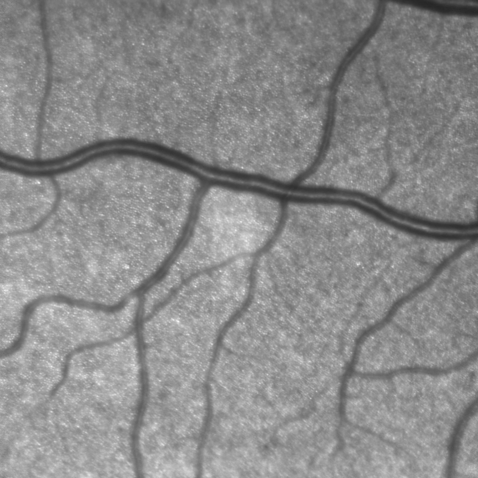
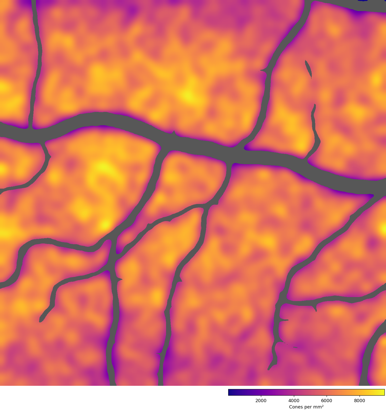
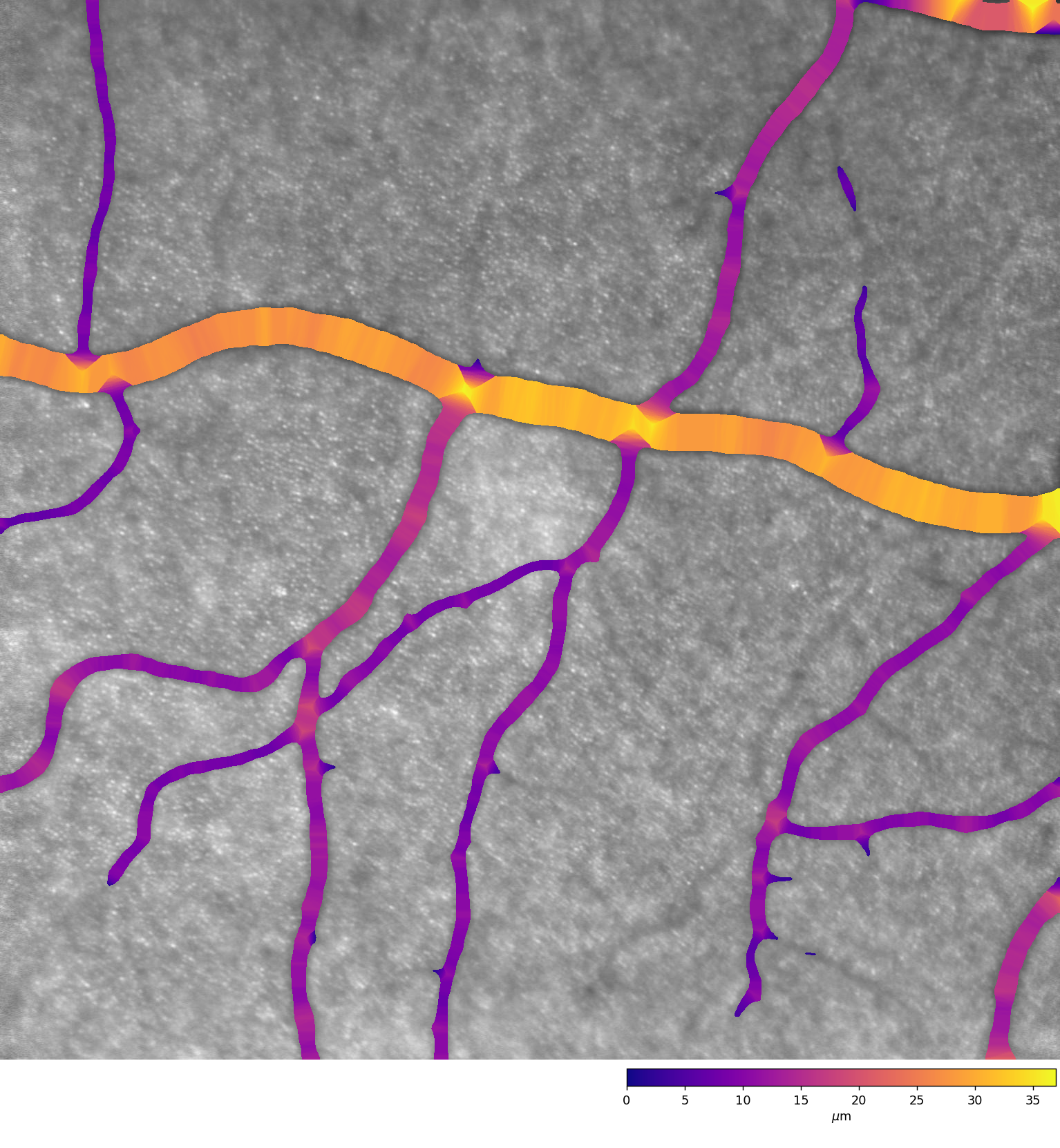
Our experience in this area includes many different imaging techniques such as optical coherence tomography (OCT), sonography (ultrasound), fluorescence angiography and retinal images employing adaptive optics (AO). In AO images, for example, the methods we have developed can be used to automatically segment the vessels and thus determine their diameter. Furthermore, individual cones can be detected, allowing cone density on the retina to be determined – a vital factor in the detection of many diseases.