Fraunhofer Institute for Computer Graphics Research IGD
Working with ultrasound image data has a long tradition at Fraunhofer IGD. As early as 1994, the first applications of 3D ultrasound imaging were developed here by Prof. Dr. Georgios Sakas. In 1997, Prof. Sakas founded the Fraunhofer spin-off MedCom, which has since been successfully involved in the processing and analysis of ultrasound image data. Meanwhile, research work and application-related projects with ultrasound image data at Fraunhofer IGD have been continuously carried on.
Automatic analysis and processing of 2D and 3D ultrasound image data
Today, the Visual Healthcare Technologies department has numerous algorithms and methods available for the automatic analysis and processing of 2D and 3D ultrasound image data.
The more recent research results include the following works:
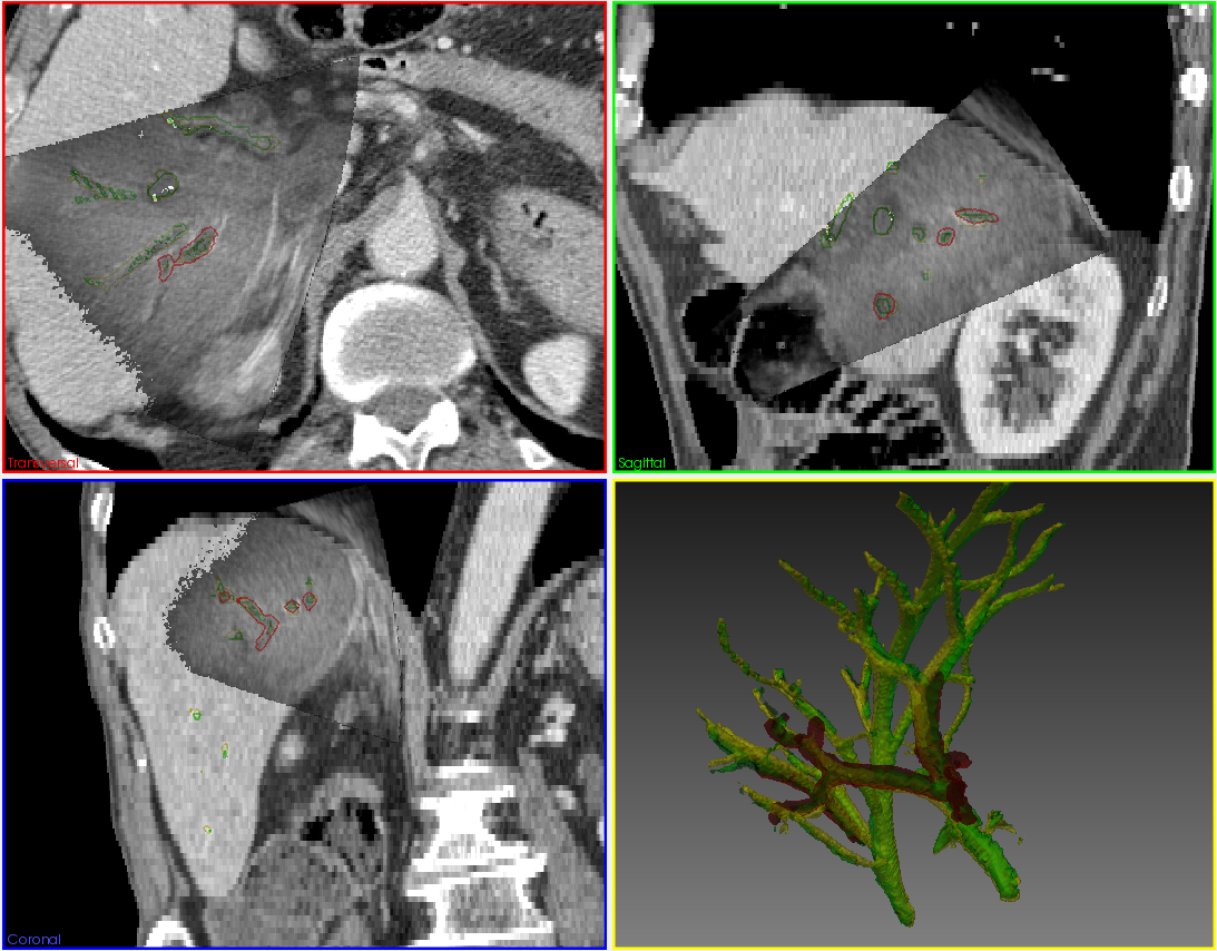
- Intraoperative registration of liver ultrasound and CT image data
- The automatic detection and segmentation of organ structures such as the liver and kidney
- Facial features of a fetus
- Ultrasound shadows e.g., of the ribs
- Free intraabdominal fluids in blunt traumas