 Fraunhofer Institute for Computer Graphics Research IGD
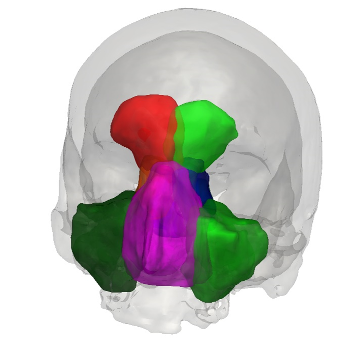
As part of the long-standing cooperation with the ENT Clinic of the University Hospital Düsseldorf, Fraunhofer IGD has also been developing solutions for the area of the paranasal sinuses for several years. This includes work on image-based automatic recognition and quantification of different pathologies (e.g., opacifications in the various paranasal sinuses). Complex health conditions that depend on a multitude of parameters are also in focus.
About 80% of the population has a deformation of the nasal septum (nasal septum deviation or septum deviation). This usually does not pose a great risk to patients. However, in severe cases, such a deformation can even cause suffocation, which is why an accurate diagnosis is of great importance. This diagnosis is made based on a multitude of parameters that can be extracted from the images.
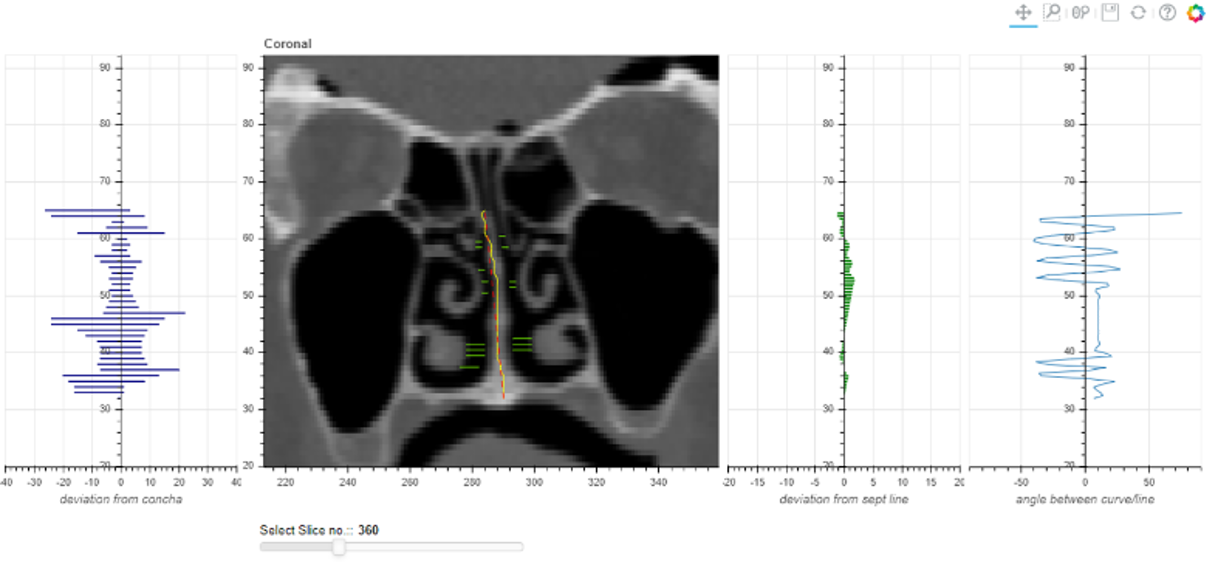
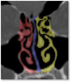
Fraunhofer IGD has developed a machine learning process that quantifies the deformation based on the automatic recognition and segmentation of several relevant structures. The segmentation framework automatically marks a multitude of structures in the CT image data: maxillary sinus, frontal sinus, sphenoid bone, conchae, and ethmoid cells on the left and right, as well as the nasal septum. For the quantification of septum deviation, the conchae and the septum play a particularly important role. The severity of the deformation depends on the angle of the nasal septum in all areas as well as on the deviation between the conchae and the undeformed midline of the septum. Through a web application, medical experts can navigate through the patient's CT dataset and read all relevant information regarding the deformation.